Reaction: MBL binds to repetitive carbohydrate structures on the surfaces of viruses, bacteria, fungi, and protozoa
- in pathway: Lectin pathway of complement activation
The MBL polypeptide chain consists of a short N-terminal cysteine-rich region, a collagen-like region comprising 19 Gly-X-Y triplets, a 34-residue hydrophobic stretch, and a C-terminal C-type lectin domain. MBL monomers associate via their cysteine-rich and collagen-like regions to form homotrimers, and these in turn associate into oligomers. The predominant oligomers found in human serum contain three (MBL-I) or four (MBL-II) homotrimers (Fujita et al. 2004, Teillet et al. 2005). Extracellular MBL oligomers circulate as complexes with MASP1/2. In the presence of Ca2+, the carbohydrate recognition domain (CRD) of MBL binds carbohydrates with 3- and 4- OH groups in the pyranose ring, such as mannose and N-acetyl-D-glucosamine. Such motifs occur on the surfaces of viruses, bacteria, fungi and protozoa. The affinity of any one MBL binding site for a carbohydrate ligand is low, but interaction between multiple binding sites on an MBL oligomer and repetitive carbohydrate motifs on a target cell surface allow high-avidity binding. The specificity of the MBL binding site (it does not bind glucose or sialic acid) and the requirement for a repeated target motif may account for the failure of MBL to bind human glycoproteins under normal conditions (Petersen et al. 2001). This reaction in particular represents the interaction of MBL with bacterial mannose repeats.
Reaction - small molecule participants:
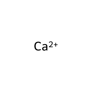
Ca2+ [extracellular region]
Reactome.org reaction link: R-HSA-166721
======
Reaction input - small molecules:
calcium(2+)
Reaction output - small molecules:
Reactome.org link: R-HSA-166721

