Reaction: Activated TAK1 mediates phosphorylation of the IKK Complex
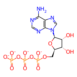
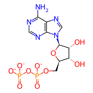
This Reactome event shows phosphorylation of IKK beta (IKBKB) by TGF-β–activated kinase 1 (TAK1), encoded by the MAP3K7 gene. TAK1 functions downstream of receptor signaling complexes in TLR, TNF-alpha and IL-1 signaling pathways (Xu & Lei 2021). TAK1 appears to be essential for IL-1-induced NF-kappa-B activation since a specific TAK1 inhibitor (5Z)-7-oxozeaenol prevents NF-kappa-B activation in human umbilical vein endothelial cells (HUVEC) (Lammel 2020); also, it prevents NF-kappa-B-mediated TNF production in human myeloid leukaemia U937 cells (Rawlins et al. 1999). TAK1 functions through assembling the TAK1 complex consisting of the coactivators TAB1 and either TAB2 or TAB3 (Shibuya et al. 1996, Sakurai et al. 2000; Xu & Lei 2021). TAB1 promotes TAK1 autophosphorylation at the kinase activation lobe (Sakurai et al. 2000; Brown et al. 2005). The TAK1 complex is regulated by polyubiquitination. The binding of TAB2 or TAB3 to polyubiquitinated TRAF6 may facilitate polyubiquitination of TAB2, -3 by TRAF6 (Ishitani et al. 2003), which in turn results in conformational changes within the TAK1 complex. TAB2 or -3 is recruited to K63-linked polyubiquitin chains of receptor interacting protein (RIP) kinase RIP1 (RIPK1) via the Zinc finger domain of TAB2 or TAB3. RIPK1 functions as an essential component of inflammatory and immune signaling pathways. Ubiquitination of RIPK1 follows the recruitment of TRADD and TRAF2 or -5 (the latter functions as the E3 ubiquitin ligase, but also cIAP1,-2 can ubiquitinate RIPK1 as a response to TNF receptor engagement (Varfolomeev et al. 2008). The IKK complex is also recruited ubiquitin (Ub) chains via its Ub binding domain. Polyubiquitin chains may function as a scaffold for higher order signaling complexes bringing the TAK1 and IKK complexes in close proximity and allowing TAK1 to phosphorylate IKBKB (IKK2) (Kanayama et al. 2004).
The alternative (non-canonical) pathway can be activated via CD40, LTßR, BAFF, RANK, and is therefore limited to cells which express these receptors. It leads to NIK-mediated phosphorylation of IKKa (IKK1, CHUK), which phosphorylates the NFKB2 (p52) precursor p100, leading to the ubiquitin-dependent degradation of its C-terminal part (processing of p100 to the mature p52 subunit) and releasing the NFKB2:RelB complex (Sun 2017). In non-stimulated cells NIK is constitutively degraded by the cIAP1/2:TRAF2:TRAF3 Ub ligase complex; following stimulation, the complex is recruited to the respective receptor comlex where cIAPs ubiquitinates TRAF3, resulting it its degradation and stabilization of NIK. NIK then phosphorylates and activates IKK1 (CHUK), leading to the NFKB2:RelB complex activation (Sun 2017). TRAF3 deubiquitylation by OTUD7B downregulates the NIK-mediated NF-kappa-B activation. (Hu et al 2013). In addition, TAK1 has been shown to interact with NIK and with IKK2, and TAK1 can be stimulated by anti-apoptotic protein, XIAP (Hofer-Warbinek et al. 2000). XIAP is an NF-kB dependent gene, therefore its expression represents a positive regulatory circuit. NIK is also involved in the classical pathway, and is activated by TAK1 in the IL-1 signalling pathway (Ninomiya-Tsui et al. 1999) and Hemophilus influenzae-induced TLR2 signalling pathway (Shuto et al. 2001).
RNA-induced liquid phase separation of SARS-CoV-2 nucleocapsid (N) protein serves as a platform to enhance the interaction between TAK1 and IKK complexes promoting NF-kappa-B-dependent inflammatory responses (Wu Y et al. 2021).


