Reaction: RIPK3 is phosphorylated
- in pathway: RIPK1-mediated regulated necrosis
Necroptosis is a form of regulated necrotic cell death mediated by interaction of receptor-interacting serine/threonine-protein kinase 1 (RIPK1) and RIPK3 via a RIP homotypic interaction motif (RHIM) domain. RIPK1:RIPK3 complex formation further potentiates kinase activation through autophosphorylation and/or transphosphorylation, propagating the pronecrotic signal. RIPK1, RIPK3 and their kinase activities were shown to be essential for necroptosis (Degterev A et al. 2008; Cho YS et al. 2009). A RIPK3 kinase-dead mutant (K50A) was found to function as a dominant negative mutant, which blocked tumor necrosis factor alpha (TNFα)-induced necrotic pathway in human colorectal adenocarcinoma HT-29 cells (He S et al. 2009). Studies in mice expressing catalytically inactive RIPK3 showed that RIPK3 D161N stimulated RIPK1-dependent apoptosis and embryonic lethality in RIPK3 D161N homozygous mice, while K51A knock in mice developed into fertile and immunocompetent adults, suggesting that the kinase activity of RIPK3 determines whether cells die by necroptosis or caspase-8-dependent apoptosis (Mandal P et al. 2014; Newton K et al. 2014; Raju S et al. 2018). Further, differentially tagged constructs of RIPK3 kinase domain (KD) were found to form dimers after their co-expression in human embryonic kidney (HEK) 293T cells, and mutation of residues at the dimer interface impaired dimerization (Raju S et al. 2018). Phosphorylation on the serine residue 227 (S227) of human RIPK3 (S231 and S232 on mouse RIPK3) is thought to mediate recruitment and activation of mixed-lineage kinase domain-like (MLKL), a crucial downstream substrate of RIPK3 in the necrosis pathway (Sun et al. 2012; Chen et al. 2013). The phosphorylation occurs in the αG helix in the C-lobe of the RIPK3 kinase, not the activation loop (Petrie EJ et al. 2019;. Consequently it remains unclear why this would be an activating event and how this would lead to MLKL interaction Although RIPK1 activation is associated with phosphorylation of the RIPK3 activation loop, most studies, however, suggest that RIPK1 does not phosphorylate RIPK3 (Cho YS et al. 2009). Rather, it is thought that active RIPK1 serves as a scaffold to enable RIPK3 to assemble into homooligomers. The precise mechanism of MLKL activation by RIPK3 is incompletely understood and may vary across species (Davies KA et al. 2020). The underlying mechanism is still debated, but the point is that RIPK3 transphosphorylation is crucial for MLKL activation (Cook WD et al. 2014; Orozco S et al. 2014; Mompean M et al. 2018).
FDA-approved anticancer drugs, including sorafenib and ponatinib, showed anti-necroptotic activity (Fauster A et al. 2015; Martens S et al. 2017; Fulda S 2018). These compounds are tyrosine kinase inhibitors (TKI) that directly targeted RIPK3 and RIPK1 and blocked their kinase activity (Fauster A et al. 2015; Martens S et al. 2017; Fulda S 2018). Pazopanib, another multi-targeting TKI, was shown to suppress necroptosis preferentially by targeting RIPK1 (Fauster A et al. 2015).
Reaction - small molecule participants:
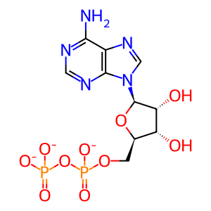
ADP [cytosol]
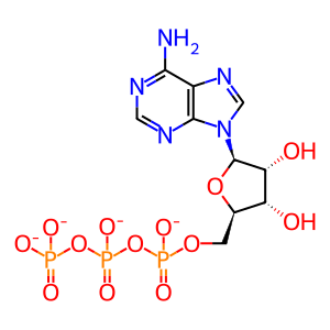
ATP [cytosol]
Reactome.org reaction link: R-HSA-5213466
======
Reaction input - small molecules:
ATP(4-)
Reaction output - small molecules:
ADP(3-)
Reactome.org link: R-HSA-5213466


