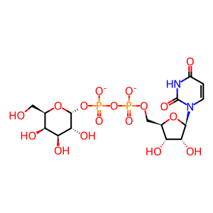
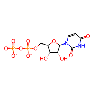
Reaction: B4GALNT2 transfers GalNAc from UDP-GalNAc to Sial-Gal-GlcNAc-Gal to form the Sd(a) antigen on UMOD
- in pathway: Asparagine N-linked glycosylation
The histo-blood group antigen Sda was discovered to be a dominant character found in more than 90 % of Caucasian red blood cells. In addition to erythrocytes, the Sda antigen is also found in other tissues and body fluids, particularly in urine of humans and other mammals. The Sda antigen shares a common minimal saccharide structure (GalNAc-beta1-4[Neu5Ac-alpha2-3]Gal-beta-) with the Cad antigen. Both antigens contain a pentasaccharide structure, the Sda antigen's structure being GalNAc-beta1-4[Neu5Ac-alpha2-3]Gal-beta1-4GlcNAc-beta1-3Gal. The last step in the biosynthesis of both Sda and Cad antigens is catalysed by beta-1,4 N-acetylgalactosaminyltransferase 2 (B4GALNT2), a Golgi membrane protein that transfers N-acetylgalactosamine (GalNAc) from UDP-GalNAc to position C4 of the Gal residue of the Neu5Ac-alpha2-3Gal-beta1 sequence (Montiel et al. 2003, Lo Presti et al. 2003, Dall'Olio et al. 2014). Both antigens can be expressed by N- or O-linked chains of glycoproteins. Tamm–Horsfall glycoprotein (THGP, aka UMOD) is a major carrier of the Sda antigen in urine (Soh et al. 1980, Serafini-Cessi & Conte 1982). After proteolytic cleavage, UMOD is secreted into the urine where it may play roles in colloid osmotic pressure, retarding passage of positively charged electrolytes, preventing urinary tract infection and modulateing formation of supersaturated salts and their crystals (Schmid et al. 2010).
Reaction - small molecule participants:
UDP [Golgi lumen]
UDP-Gal [Golgi lumen]
Reactome.org reaction link: R-HSA-8855954
======
Reaction input - small molecules:
UDP-alpha-D-galactose(2-)
Reaction output - small molecules:
UDP(3-)
Reactome.org link: R-HSA-8855954


