Binding of TNFα to TNF receptor 1 (TNFR1) induces the sequential formation of several signaling complexes, namely complex I and complex IIa/b (Micheau O and Tschopp J 2003; Walczak H 2011; Yuan J et al. 2019). These complexes support either cell survival (complex I) or cell death (complex II). The dynamic assembly of these complexes is tightly regulated by proteolysis, ubiquitination and phosphorylation of receptor-interacting serine/threonine protein kinase 1 (RIPK1) and other components of the TNFα signaling pathway (reviewed in Yuan J et al. 2019; Varfolomeev E & Vucic D 2022). RIPK1 functions as a key regulator of both cell survival and cell death (reviewed in Ju E et al. 2022). Deubiquitination of RIPK1 leads to the activation of RIPK1 kinase activity, which promotes autophosphorylation of RIPK1 and RIPK1 kinase-dependent cell death (reviewed in Ju E et al. 2022). The kinase activity of RIPK1 is regulated by phosphorylation (Lafont E et al. 2018; Dondelinger Y et al. 2019; reviewed in Delanghe T et al. 2020; Ju E et al. 2022). The components of I-kappa-B kinase complex, namely inhibitor of nuclear factor kappa B subunit α (IKKα or CHUK) and subunit β (IKKβ or IKBKB), can directly phosphorylate RIPK1 within the membrane-bound TNFR1 signaling complex (complex I) (Dondelinger Y et al. 2019; Delanghe T et al. 2020). In addition, TANK binding kinase 1 (TBK1) and IKKε (IKBKE) are recruited to complex I to phosphorylate RIPK1 independently from IKKα/IKKβ. These phosphorylation events suppress activation of RIPK1 within complex I, preventing subsequent formation of the death-inducing complex II (Lafont E et al. 2018; reviewed in Delanghe T et al. 2020). In the cytosol, RIPK1 is phosphorylated by MAPK-activated protein kinase 2 (MAPKAPK2 or MK2) (Jaco I et al. 2017; Dondelinger Y et al. 2017; Menon MB et al. 2017). MAPKAPK2 (MK2) deficiency sensitized human and mouse cells to TNF-induced RIPK1-dependent cell death (Jaco I et al. 2017; Dondelinger Y et al. 2017; Menon MB et al. 2017; Chen IT et al. 2021). In the mouse model of TNFα-induced systemic inflammation, MAPKAPK2 (MK2)-deficient mice exhibited significantly increased inflammatory response, cellular damage and mortality compared to wild type controls following treatment with TNFα (Vandendriessche B et al. 2014; Dondelinger Y et al. 2017). Mass spectrometry (MS)-based phosphoproteomics using mouse LPS-treated bone marrow-derived macrophage (BMDM) and stress-stimulated embryonic fibroblasts (MEF) identified RIPK1 as a direct substrate of MAPKAPK2 (Menon MB et al. 2017). Immunoblotting and mutagenesis analysis further confirmed MAPKAPK2 (MK2)-dependent phosphorylation of RIPK1 in TNF-stimulated mouse and human cells (Jaco I et al. 2017; Dondelinger Y et al. 2017; Menon MB et al. 2017). MAPKAPK2-mediated phosphorylation of RIPK1 inhibited the ability of RIPK1 to bind to FADD in mouse cells (Jaco I et al. 2017; Dondelinger Y et al. 2017; Menon MB et al. 2017). MAPKAPK2 was shown to phosphorylate mouse RIPK1 at serine 321 (S321) and S336 (equivalent to S320 and S335 in human RIPK1) (Jaco I et al. 2017; Dondelinger Y et al. 2017; Menon MB et al. 2017). Other protein kinases such as transforming growth factor β-activated kinase 1 (TAK1) or IKKα/IKKβ may also regulate phosphorylation of RIPK1 at these phospho-acceptor sites (Krishnan RK et al. 2015; Mohideen F et al. 2017; Geng J et al. 2017). Functional analysis revealed that phosphorylation of human RIPK1 at S320 negatively regulates RIPK1 autophosphorylation at S166, which is essential for the RIPK1 kinase activity and subsequent induction of cell death signaling (Mohideen F et al. 2017; Du J et al. 2021). While phosphorylation of RIPK1 at S320 had no effect on TNFα-induced NF-κappa-B signaling, it limited RIPK1 kinase activation and association with FADD and CASP8 in human cells (Mohideen F et al. 2017). Similar results were obtained for S321/S336 of mouse RIPK1 (Geng J et al. 2017; Dondelinger Y et al. 2017; Menon MB et al. 2017; Jaco I et al. 2017). These data suggest that MAPKAPK2-mediated phosphorylation of RIPK1 at S320 limits TNFα-induced RIPK1-dependent cell death. Besides TNFR1, MAPKAPK2 regulates RIPK1 activity downstream of Toll-like receptor 4 (TLR4) upon LPS stimulation (Jaco I et al. 2017; Menon MB et al 2017). Moreover, MAPKAPK2 controls inflammation by regulating expression of inflammatory cytokines such as TNFα, IL-1β and IL-6 (reviewed in Morgan D et al. 2022).
Upon TNFa stimulation, MAPKAPK2 (MK2) is activated by p38 MAPK downstream of TAK1, which is recruited to and activated in the TNFR1 signaling complex (Jaco I et al. 2017). Activated TAK1 mediates TNF-induced NF-kappa-B and MAPK signaling pathways. Pathogenic bacteria developed strategies to suppress host immune responses by blocking activation of NF-kappa-B and MAPK signaling pathways (Philip NH et al. 2014; He C et al. 2017; Orning P et al. 2018; Malireddi RKS et al. 2020). For example, the cytotoxicity of pathogenic Yersinia species is caused by Yersinia outer protein YopJ secreted in Y. pestis and Y. pseudotuberculosis and YopP secreted by Y. enterocolitica. The acetyltransferase activity of YopJ/P catalyses O-acetylation of specific serine and/or threonine residues in the activation loop of protein kinases. YopJ/P-mediated acetylation of TAK1 suppresses the catalytic activity of TAK1 and thus preventing production of pro-inflammatory cytokines in human and mouse cells (Thiefes A et al. 2006; Paquette N et al. 2012). Further, YopP was shown to suppress TAK1/p38 MAPK/MK2 signaling while inducing activation of RIPK1 in Y. enterocolitica-infected mouse J774A.1 macrophages (Menon MB et al. 2017). These data suggest that Yersinia-mediated inactivation of TAK1 leads to induction of RIPK1-dependent cell death, which contributes to host defense against pathogens (Weng D et al. 2014; Philip NH et al. 2014; Menon MB et al. 2017; Peterson LW et al. 2017; Orning P et al. 2018).
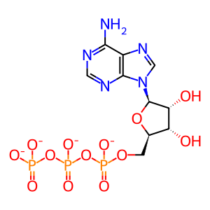
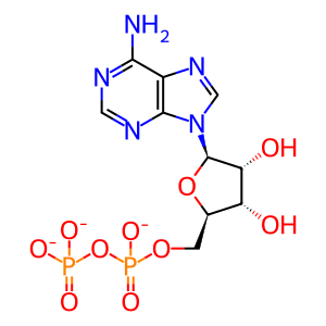
This Reactome event shows MAPKAPK2 (MK2)-mediated phosphorylation of cytosolic RIPK1 at S320 in response to TNFα stimulation.


