Reaction: TBK1, IKBKE phosphorylate RIPK1 at T189
- in pathway: Regulation of TNFR1 signaling
Binding of TNFα to TNF receptor 1 (TNFR1) induces either cell survival or cell death through the sequential formation of several signaling complexes, namely complex-I, IIa and IIb (Micheau O and Tschopp J 2003; Walczak H 2011). The dynamic assembly of these complexes is tightly regulated by proteolysis, ubiquitination, deubiquitination and phosphorylation of receptor-interacting serine/threonine protein kinase 1 (RIPK1) and other components of the TNFα signaling pathway. The rapidly forming complex-I (the TNFR1 signaling complex) is assembled at the receptor’s cytoplasmic tail and consists of TNFR1, TNFR1-associated death domain (TRADD), TNF receptor associated factor-2 (TRAF2) and RIPK1 (Micheau O and Tschopp J 2003). The activated TNFR1 signaling complex (complex-I) recruits several E3 ubiquitin (Ub) ligases, such as cIAP1/2 cellular inhibitor of apoptosis (BIRC2, BIRC3), mind bomb 2 (MIB2) or linear ubiquitin chain assembly complex (LUBAC) (Micheau O and Tschopp J 2003; Feltham R et al. 2018; Yuan J et al. 2019). K63-linked ubiquitination by BIRC2, BIRC3 and MIB2 as well as LUBAC-mediated Met1-linked linear ubiquitination of RIPK1 and other complex components stabilize the membrane-bound pro-survival TNFR1 signaling complex, while suppressing the formation of the cytosolic death-inducing complex IIa (TRADD:TRAF2:RIPK1:FADD:CASP8) and IIb (RIPK1:RIPK3:MLKL). The catalytic activity of LUBAC also enables recruitment of TRAF-associated NF-κB activator (TANK) binding kinase 1 (TBK1) and inhibitor-kappa-B kinase (IKK) epsilon (IKKε or IKBKE) to the TNFR1 signaling complex (Lafont E et al. 2018; Xu D et al. 2018). LUBAC is composed of the HOIP, HOIL-1L, and SHARPIN subunits. HOIP deficiency strongly diminished recruitment of TBK1 or IKBKE to the complex-I in human cervical carcinoma epithelial HeLa cells and lung carcinoma epithelial A549 cells (Lafont E et al. 2018). Western blot analysis revealed that TBK1 and IKBKE are recruited to and strongly phosphorylated within the TNFR1 complex in various TNFα-stimulated human cell lines including A549, keratinocyte HaCaT, monocytic U937 and THP-1 cells (Lafont E et al. 2018). Similarly, TBK1 was found to associate with components of the complex-I in the LUBAC-dependent manner in mouse cells (Xu D et al. 2018). IKBKE, but not TBK1, was detected within the TNFR1 complex in human telomerase reverse transcriptase (TERT)-immortalized dermal fibroblasts derived from patients with TBK1 deficiency as a result of loss-of-function mutation at W619* (Taft J et al. 2021). The enzymatic activity of TBK1/IKBKE is initiated by phosphorylation at S172 located in the T loop of the TBK1 and IKKε kinase domains (Shimada T et al. 1999; Kishore N et al. 2002; Gu L et al. 2013). Activated TBK1 and IKBKE are known to trigger phosphorylation of interferon regulatory factor 3 (IRF3) and IRF7 and subsequent expression of type I interferons (IFNs; IFN-α/β) downstream of pattern recognition receptors, such as Toll-like receptors 3 and 4 (TLR3, TLR4), cGAS/STING and RIG-I-like receptors (Fitzgerald KA et al. 2003; Fang R et al. 2017). While TBK1 or IKBKE showed limited effect on TNF-induced gene expression in human A549 and mouse embryonic fibroblasts (MEF) (Lafont E et al. 2018), both kinases prevented TNFα-induced cell death by suppressing RIPK1 activation in human and mouse cells (Lafont E et al. 2018; Xu D et al. 2018; Taft J et al. 2021). Mechanistically, TBK1 and/or IKBKE directly phosphorylate RIPK1 within the TNFR1 signaling complex to prevent RIPK1 kinase-dependent cell death signaling downstream of TNFR1 (Lafont E et al. 2018; Xu D et al. 2018). Mass spectrometry analysis suggests that human RIPK1 is phosphorylated by TBK1 at T189 (Xu D et al. 2018). Using a phospho-specific antibody, T189 of RIPK1 was detected as a phosphorylation site upon incubation with TBK1 in in vitro kinase assay and upon co-expression of tagged proteins in human embryonic kidney HEK293T cells. This phosphorylation was inhibited by TBK1/IKBKE inhibitor MRT67307 (Xu D et al. 2018). Phosphorylation of endogenous RIPK1 at T189 was detected in TNF-stimulated Jurkat cells (Xu D et al. 2018). Others reported that TBK1 or IKBKE phosphorylate RIPK1 at multiple residues (Lafont E et al. 2018). Further, TBK1 or IKBKE deficiency enhanced TNF-induced autophosphorylation of RIPK1, association of RIPK3 with caspase-8:FADD and levels of phosphorylated MLKL in human A549 and HeLa cells (Lafont E et al. 2018). Similar findings were reported for mouse cells (Lafont E et al. 2018; Xu D et al. 2018). In vivo studies showed that the embryonic lethality of Tbk1−/− mice was caused by TNFα-stimulated hyperactivation of RIPK1 kinase activity (Xu D et al. 2018). Moreover, chronic systemic autoinflammation in patients carrying homozygous loss-of-function mutations in TBK1 (W619*, R440* and Y212D) is associated with enhanced TNFα-induced RIPK1-dependent cell death (Taft J et al. 2021). In addition, TBK1-mediated phosphorylation of CYLD may also control the TNFR1 signaling pathway by regulating the ubiquitination status of RIPK1 (Xu X et al. 2020; Taft J et al. 2021). These data suggest that TBK1 and IKBKE downregulate RIPK1 auto-phosphorylation within the complex-I, thereby preventing TNFα- induced RIPK1-kinase-activity-dependent cell death.
This Reactome event describes TBK1/IKBKE-mediated phosphorylation of RIPK1 at T189 within the TNFR1 signaling complex.
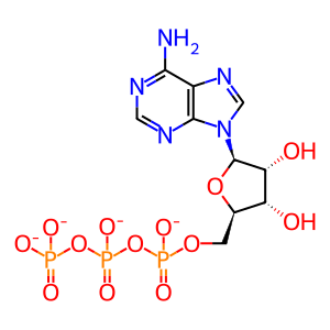
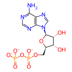
Reaction - small molecule participants:
ADP [cytosol]
ATP [cytosol]
Reactome.org reaction link: R-HSA-9817397
======
Reaction input - small molecules:
ATP(4-)
Reaction output - small molecules:
ADP(3-)
Reactome.org link: R-HSA-9817397


