Reaction: Collagen prolyl 3-hydroxylase converts 4-Hyp collagen to 3,4-Hyp collagen
- in pathway: Collagen biosynthesis and modifying enzymes
Collagen contains (2S,3S)-3-hydroxyproline (3-Hyp), though much less abundantly than 4-Hyp (Rhodes and Miller 1978). The 3-Hyp content of collagen is much more variable than that of 4-Hyp, varying between collagen types, tissues, developmental stages and pathological states (Kivirikko et al. 1992). It is more prevalent in type IV and V collagens at 10-15 3-Hyp residues (Bentz et al. 1983) than in Type I-III fibrillar collagens which have a single 3-Hyp residue per chain; the alpha-1 chain of type I collagen has 3-Hyp at residue 986 (Fietzek et al. 1972, Marini et al. 2007). 3-Hyp is formed from Pro in the Xaa position of Xaa-Hyp-Gly triplets (Gryder et al. 1975, Kivirikko et al. 1992). It is likely that 4-Hyp is a requirement at the second position of the triplet as 4-Hyp rich substrates are more active than 4-Hyp poor (Adams & Frank 1980). 3-Hyp has a modest effect on triple-helix stability (Jenkins et al. 2003; Mizuno et al. 2008). 3-Hyp may adjust the stability of basement membranes to enable formation of the meshwork structure, or serve as a ligand for other proteins. It is suggested to have a role in the self-assembly of collagen supramolecular structures (Weis et al. 2010).
3-Hyp is formed by prolyl 3-hydroxylase (P3H; EC 1.14.11.7), which has 3 isoforms in vertebrates. All contain an ER-retention signal but vary in their tissue expression (Vranka et al. 2009). P3H can hydroxylate prolines that precede 4-Hyp residues (Tryggvason et al. 1976) but not those that precede an unhydroxylated proline (Kivirikko & Myllla 1982, Myllyharju 2005). Like P4H, P3H requires molecular oxygen, alpha-ketoglutarate, iron(II), and ascorbate for activity. P3H1 is homologous to mammalian leprecan or growth suppressor 1 (Gros1), and forms a 3-prolyl hydroxylation complex with cartilage-associated protein (CRTAP) and a peptidyl-prolyl cis-trans isomerase, cyclophilin B (CypB), which is encoded by the PPIB gene (Vranka et al. 2004). Lack of 3-Hyp in Type I and II collagens leads to an osteogenesis imperfecta (OI)-like disease, as demonstrated by CRTAP and PPIB knock-out mice (Morello et al. 2006, Choi et al. 2009) and by mutations in human LEPRE1 (which encodes P3H1), CRTAP, and PPIB (Barnes et al. 2006, Cabral et al. 2007, van Dijk et al. 2009). The P3H1/CRTAP/CypB complex has also been shown to have chaperone activity (Ishikawa et al. 2009). P3H2 hydroxylates peptides derived from Type IV collagen more efficiently than Type I peptides and is localized to tissues that are rich in basement membrane (Tiainen et al. 2008). The effect of prolyl 3-hydroxylation on basement membrane collagens remains unknown.
In this generalized reaction, all collagen subtypes are represented as having one 3-Hyp residue.
Reaction - small molecule participants:
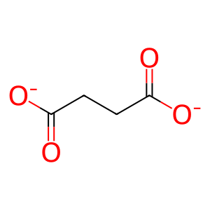
SUCCA [endoplasmic reticulum lumen]
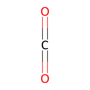
CO2 [endoplasmic reticulum lumen]
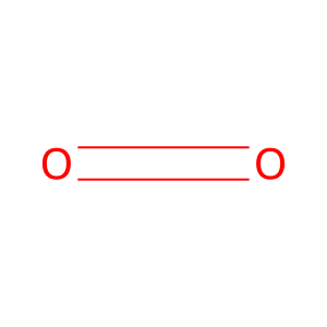
O2 [endoplasmic reticulum lumen]
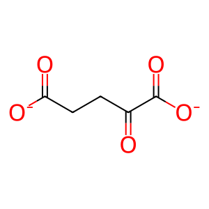
2OG [endoplasmic reticulum lumen]
Reactome.org reaction link: R-HSA-1980233
======
Reaction input - small molecules:
dioxygen
2-oxoglutarate(2-)
Reaction output - small molecules:
succinate(2-)
carbon dioxide
Reactome.org link: R-HSA-1980233


